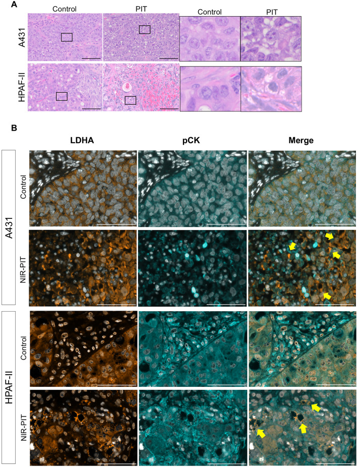
Fig. 5.
Histological changes after in vivo TF-targeted NIR-PIT. Tumor tissue histology was examined 24 h after NIR-PIT. A, H&E staining of A431 and HPAF-II tumors after NIR-PIT (images, × 200; scale bar, 100 μm). Insets are enlarged and displayed in the right panels. B, Immunohistochemical evaluation of lactate dehydrogenase A (LDHA) expression in A431 and HPAF-II tumors 24 h after NIR-PIT. Representative pictures of LDHA expression (images; × 200; scale bar, 100 μm). The inset shows examples of LDHA leakage into the extracellular space, which suggests necrotic cell death (yellow-filled arrow). Antibody staining of LDHA and pan-cytokeratin (pCK) is shown in orange and cyan, respectively. Nuclei are stained with DAPI and shown in white

