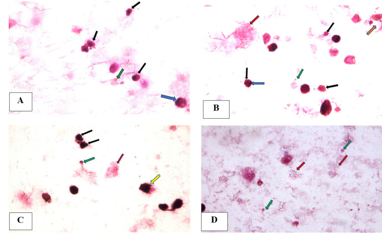
Figure 7.
(A) A photomicrograph of tongue carcinoma cells after 24-hour treatment with piperine featuring apoptotic cells with apoptotic nuclei (black arrows), apoptotic body (green arrow) and peripheral condensation of chromatin (blue arrow), (B) A photomicrograph of tongue carcinoma cells after 24-hour treatment with cisplatin showing apoptotic cells (black arrows) with peripheral condensation of chromatin (blue arrow) , nuclear fragmentation (orange arrow) and apoptotic body (green arrow) and necrotic cell (red arrow), (C) (D) A photomicrograph of tongue carcinoma cells after 24-hour treatment with combination of piperine and cisplatin showing apoptotic cells with apoptotic nuclei (black arrows), apoptotic bodies (green arrows), membrane blebbing (yellow arrow) and necrotic debris (red arrow) (H & E, x1000 oil).

