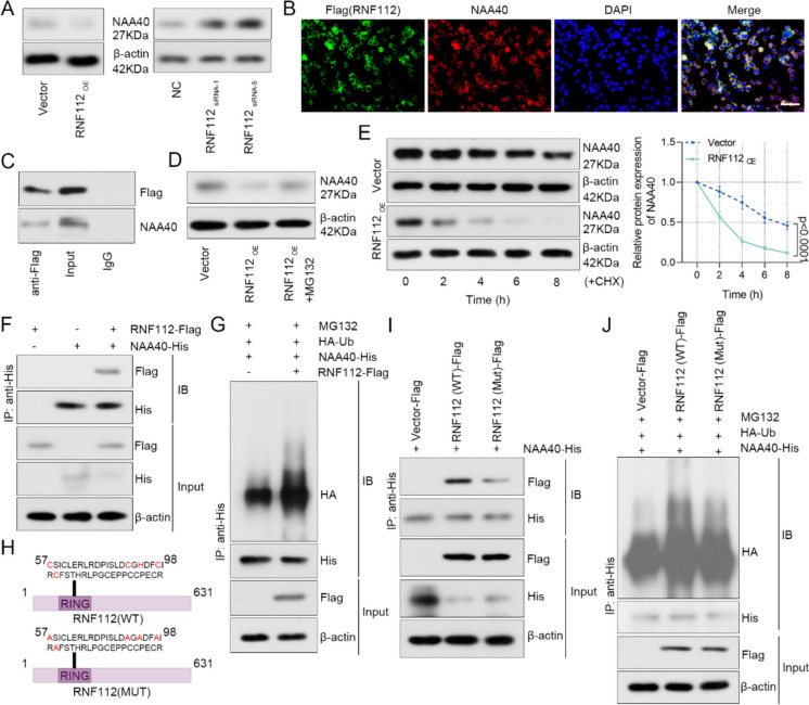
Fig. 8.
RNF112 interacted with NAA40 and enhanced its ubiquitination degradation. A SW620 cells were transfected with RNF112 overexpression plasmid or siRNA targeting RNF112 sequences. The expression of NAA40 was detected by western blot 48 h after transfection. B The co-localization of Flag (RNF112) and NAA40 in SW620 cells was detected by immunofluorescence double staining. (Scale bar = 50 µm). C Co-IP was used to detect the binding of Flag (RNF112) and NAA40. D The expression of NAA40 was detected by western blot after SW620 cells were treated with 10 µM MG132 for 8 h. E After 100 µg/ml CHX treatment for 0, 2, 4, 6 and 8 h, the expression of NAA40 was tested by western blot, and the degradation rate of NAA40 protein was calculated. F HEK293T cells were transfected with NAA40 vector (with His tag) and RNF112 vector (with Flag tag) alone or co-transfected. After 48 h of transfection, co-IP was used to detect the binding of RNF112 and NAA40. G HEK293T cells were transfected with NAA40 vector (with His tag), RNF112 vector (with Flag tag) and Ub vector (with HA tag). After transfection for 48 h and treatment with 10 µM MG132 for 8 h, co-IP was used to test the ubiquitination levels in the cells. H Schematic diagram of RNF112-Mut lacking E3 ubiquitin ligase activity. I HEK293T cells were transfected with NAA40 vector (with His tag) and RNF112-WT vector (with Flag tag) or RNF112-Mut vector (with Flag tag). After 48 h of transfection, co-IP was used to detect the binding of RNF112 and NAA40. J HEK293T cells were transfected with NAA40 vector (with His tag), RNF112-WT (with Flag tag) or RNF112-Mut (with Flag tag) and Ub vector (with HA tag). After transfection for 48 h and treatment with 10 µM MG132 for 8 h, co-IP was used to test the ubiquitination levels in the cells. (n = 3). p < 0.0001

