Figure 3.
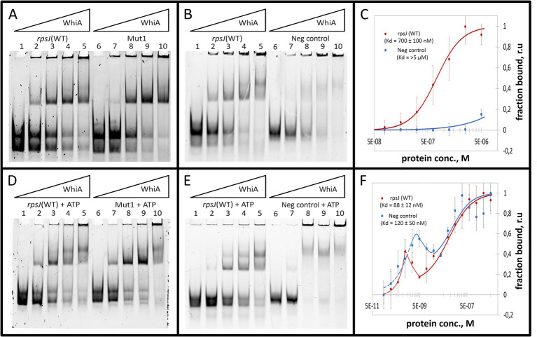
Mgal-WhiA Kd determination in the absence and in the presence of 1 mM ATP. (A) Titration of the oligonucleotide with wild-type Mgal-WhiA binding site from the rpsJ operon promoter (rpsJ WT) in parallel with the oligonucleotide with mutated aux motif (Mut1). The bands are schematically labeled on the left (from bottom to top): labeled ss-oligonucleotide, ds-oligonucleotide, the monomeric complex, the dimeric complex. Lanes: 1 – free rpsJ WT oligonucleotide 250 nM (same concentration for all lanes), 2 – 360 nM Mgal-WhiA, 3 – 720 nM Mgal-WhiA, 4 – 1,080 nM Mgal-WhiA, 5 – 1,435 nM Mgal-WhiA, 6 – free Mut1 oligonucleotide 250 nM, 7 – 360 nM Mgal-WhiA, 8 – 720 nM, 9 – 1,080 nM Mgal-WhiA, 10 – 1,435 nM Mgal-WhiA. (B) Titration of the oligonucleotide with wild-type Mgal-WhiA binding site from the rpsJ operon promoter (rpsJ WT) in parallel with the negative control oligonucleotide (Neg. control). Lanes: 1 – free rpsJ WT oligonucleotide 250 nM, 2 – 360 nM Mgal-WhiA, 3 – 720 nM, 4 – 1,080 nM Mgal-WhiA, 5 – 1,435 nM Mgal-WhiA, 6 – free negative control oligonucleotide 250 nM, 7 – 360 nM Mgal-WhiA, 8 – 720 nM, 9 – 1,080 nM Mgal-WhiA, 10 – 1,435 nM Mgal-WhiA. (C) Determination of Mgal-WhiA binding constant by microscale thermophoresis (MST) for wild-type binding site (rpsJ WT) and negative control (Neg. control) oligonucleotides. (D) Titration of the oligonucleotide with wild-type Mgal-WhiA binding site from the rpsJ operon promoter (rpsJ WT) in parallel with the oligonucleotide with mutated aux motif (Mut1) in the presence of 1 mM ATP. Lanes: 1 – free rpsJ WT oligonucleotide 250 nM (same concentration for all lanes), 2 – 360 nM Mgal-WhiA, 3 – 720 nM, 4 – 1,080 nM Mgal-WhiA, 5 – 1,435 nM Mgal-WhiA, 6 – free Mut1 oligonucleotide 250 nM, 7 – 360 nM Mgal-WhiA, 8 – 720 nM, 9 – 1,080 nM Mgal-WhiA, 10 – 1,435 nM Mgal-WhiA. (E) Titration of the oligonucleotide with wild-type Mgal-WhiA binding site from the rpsJ operon promoter (rpsJ WT) in parallel with the negative control oligonucleotide (Neg. control) in the presence of 1 mM ATP. Lanes: 1 – free rpsJ WT oligonucleotide 250 nM, 2 – 360 nM Mgal-WhiA, 3 – 720 nM, 4 – 1,080 nM Mgal-WhiA, 5 – 1,435 nM Mgal-WhiA, 6 – free negative control oligonucleotide 250 nM, 7 – 360 nM Mgal-WhiA, 8 – 720 nM, 9 – 1,080 nM Mgal-WhiA, 10 – 1,435 nM Mgal-WhiA. (F) Determination of Mgal-WhiA binding constant by microscale thermophoresis (MST) for the wild-type binding site (rpsJ WT) and negative control (Neg. control) in the presence of 1 mM ATP.

