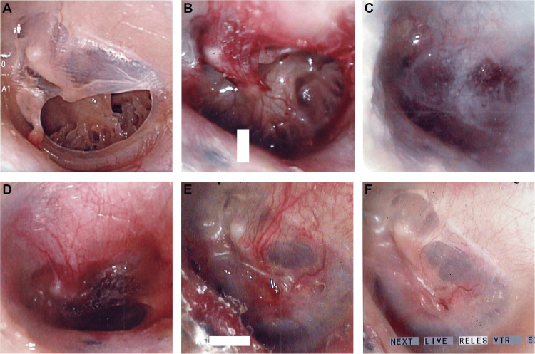
Figure 1. Images from a 30-year-old female with left TMP.
(A) Grade II TMP with calcification.
(B) The edge of the TMP was mechanically disrupted, including the calcification.
(C) A gelatin sponge immersed in bFGF was inserted into the perforation, followed by dripping fibrin glue over the sponge to seal the surgical site.
(D) One week after surgery.
(E) One and a half months after surgery. The TMP was closed.
(F) Two months after surgery. The regenerated TM resembled a natural TM.
TMP, tympanic membrane perforation; TM, tympanic membrane

