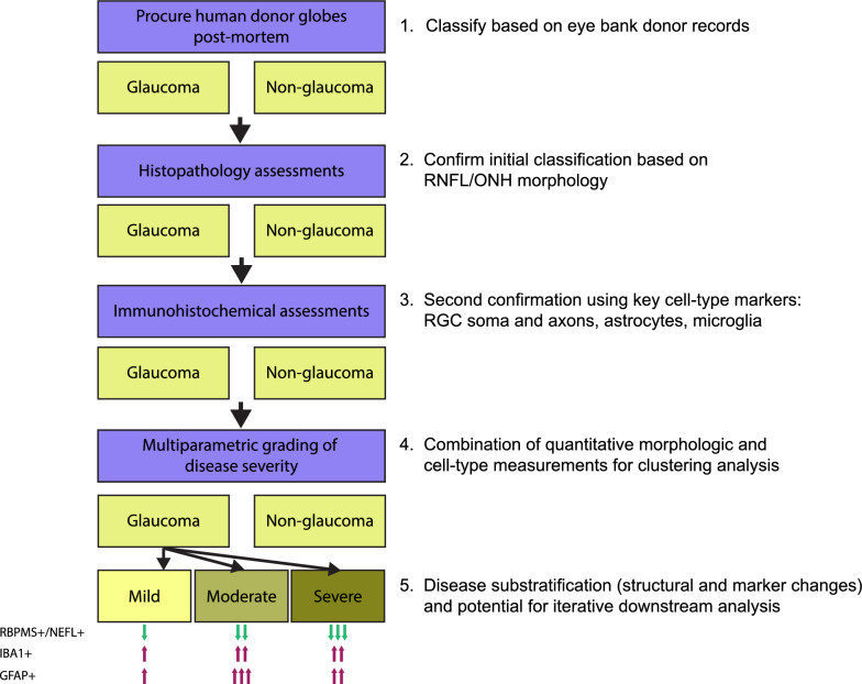
Fig. 10.
Schematic of donor eye grading workflow. Conceptual outline of steps in acquisition and grading of post-mortem donor eyes, accompanied by qualitative descriptions of protein marker stain changes from non-glaucoma samples in each severity grade. Note that the evaluation of protein markers is confined to the temporal retina. One arrow indicates a mild/minimal change, two arrows indicate a moderate change, and three arrows indicate a severe/maximal change. The predominant/distinguishing features of each grade are: mild – pre-mortem history of glaucoma; moderate – maximal GFAP positivity in temporal neural retina; severe – maximal loss of RBPMS and NEFL positivity in temporal retina

