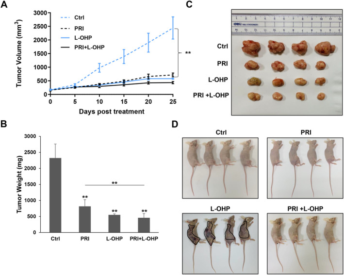
FIGURE 8.

The combination of PRIMA‐1met and L‐OHP efficiently suppressed CRC xenograft growth with low toxicity. (A) DLD‐1 (p53‐mutant) cells were inoculated into BALB/c mice (via subcutaneous injection) to establish a CRC tumor model. The detailed treatment regime and group were indicated in the Materials and Methods section. The tumor volume was measured by caliper. The tumor growth curves were constructed according to the average tumor volume of each group ± SD (mm3). **p < 0.01. (B) At the end of experiments, mice were sacrificed, and then the tumor weight was taken as shown in the bar graph. Data were reported as the mean ± SD and were analyzed by Student's t‐test; n = 4 mice per group. **p < 0.01. (C) Images of DLD‐1 tumor xenografts from mice treated with vehicle control and different drugs after dissection were shown. (D) Images of whole bodies of xenograft mice treated with vehicle control and different drugs were displayed. Petechiae and ecchymoses caused by abnormal clotting and bleeding were presented in L‐OHP‐treated mice (indicated by black shapes), but not in combination‐treated mice. Ctrl: vehicle control; PRI: PRIMA‐1met; L‐OHP: oxaliplatin.
