Figure 1.

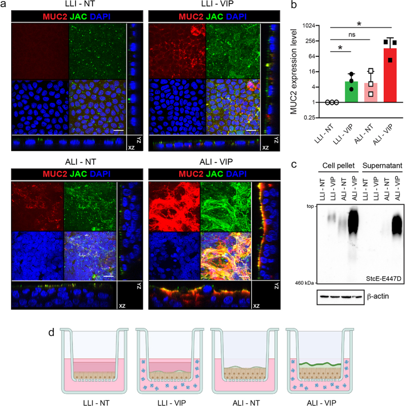
Combining air–liquid interface (ALI) and VIP treatment stimulates mucus production in caco-2 transwell cultures.
(a) Confocal microscopy of confluent low-glucose-adapted Caco-2 cultures grown on Transwell membranes in LLI and ALI conditions, not treated (NT) or treated with VIP. Cultures are stained for Jacalin (JAC, green), MUC2 (red), and nuclei (DAPI, blue). Maximum projections and orthogonal views are shown for the separate and combined channels. The white scale bar represents 20 μm. (b) qRT-PCR analysis of MUC2 expression in LLI-NT, LLI-VIP, ALI-NT, and ALI-VIP normalized to expression of ACTB. The graph displays the mRNA expression level relative to LLI-NT. Each dot represents a biological replicate. Statistical analysis was performed by a one-sample t-test. *p < .05 (c) Immunoblot analysis of mucin expression in the pellets and supernatant fractions of Caco-2 cells grown in LLI-NT, LLI-VIP, ALI-NT, and ALI-VIP conditions, visualized with the StcE-E447D probe which recognizes mucin-type O-glycosylated proteins. The top of the image corresponds to the loading slots of the agarose gel. The bottom of the image corresponds to the marker band of 460 kDa. (d) Schematic overview of Transwells containing Caco-2 cultures under LLI-NT, LLI-VIP, ALI-NT, and ALI-VIP conditions. LLI conditions contain medium in the top compartment, while the medium is removed in the ALI culture. Under LLI conditions, cultures are single-layered, while they become multilayered in ALI culture. VIP (indicated in blue) is added to the basolateral compartment. The highest mucus production is observed in the ALI-VIP combined conditions, which is the only condition showing clear apical mucus secretion (mucus layer indicated in green).
