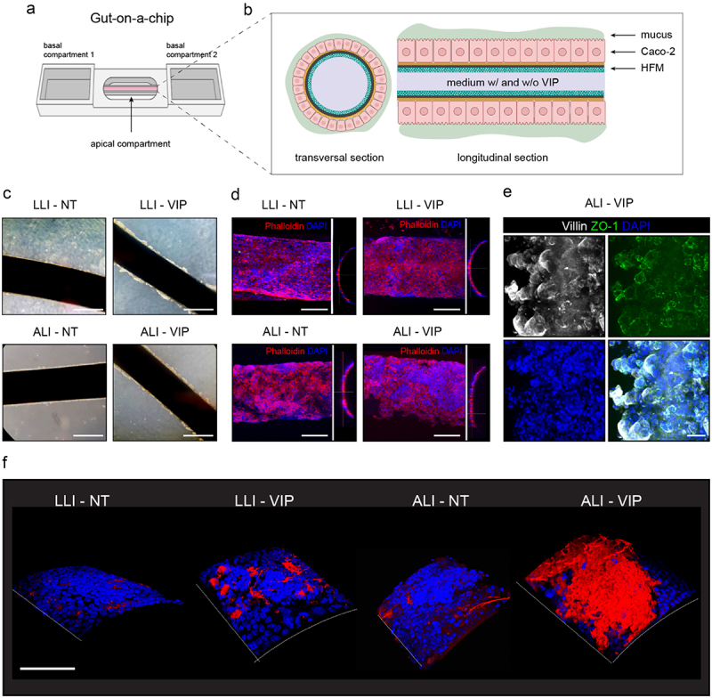
Figure 2.

ALI-VIP conditions induce MUC2 production in a gut-on-a-chip model.
(a) Gut-on-a-chip model consisting of a hollow fiber membrane (HFM) coated with Caco-2 cells mounted on a chip with 2 side reservoirs (the basolateral compartments) and one middle reservoir (the apical compartment). Cells are grown on the surface of the HFM and can be perfused basally through the fiber and apically via the inner compartment. (b) Schematic representation of the Caco-2 coated HFMs with formation of a secreted mucus layer. (c) HFMs coated with Caco-2 cells grown under LLI-NT, LLI-VIP, ALI-NT, and ALI-VIP conditions were captured using light microscopy. The white scale bar represents 500 μm. (d) Fluorescent confocal microscopy images of HFMs coated with Caco-2 cells grown under LLI-NT, LLI-VIP, ALI-NT, and ALI-VIP conditions stained for actin with phalloidin and nuclei with DAPI. The white scale bar represents 200 μm. (e) Fluorescent confocal microscopy of ALI-VIP HFM stained for villin, ZO-1, and DAPI. The white scale bar represents 50 μm. (f) 3-dimensional images of HFMs with Caco-2 cells stained for MUC2 (red) and nuclei (DAPI, blue). The white scale bar represents 200 μm.
