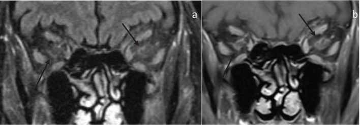
Figure 2.
80-year-old presenting with acute left eye vision loss (OS) with jaw claudication, headache, and weight loss. A fundoscopic exam demonstrated left optic disc edema. Subsequent biopsy confirmed GCA. (A) Coronal fat-saturated FLAIR demonstrates FLAIR hyperintensity along the bilateral intraorbital optic nerves sheath (arrows), suggestive of optic perineuritis. (B) The coronal fat-saturated post-contrast sequence demonstrates enhancement of the bilateral intraorbital optic nerve sheath suggestive of perineuritis (arrows).

