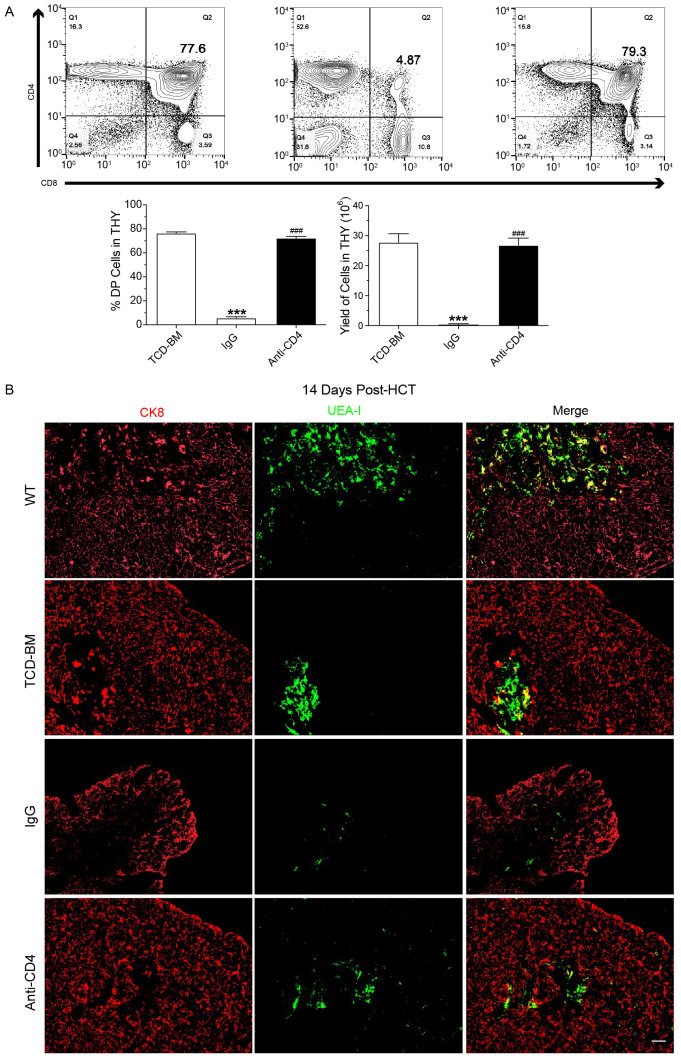
Figure 2.
Depletion of donor CD4+ T cells promotes recovery of CD4+CD8+ thymocytes and medullary thymic epithelial cells. (A) Representative FACS images (top) at day 60 post-HCT and mean ± SEM of percentage and yield (bottom) of CD8+CD4+ cells in the thymus of TCD-BM, IgG and Anti-CD4 groups. n=8 per group. (B) Representative immunostaining images of CK8 and UEA-I in the thymus of wild type mice (WT) TCD-BM, IgG and Anti-CD4 groups at day14 post-HCT. n=4 per group. Scale bar: 50 μm. The statistical significance was performed according to one-way ANOVA followed by Tukey’s post hoc. ***p < 0.001 versus TCM-BM. ###p < 0.001 versus IgG.

