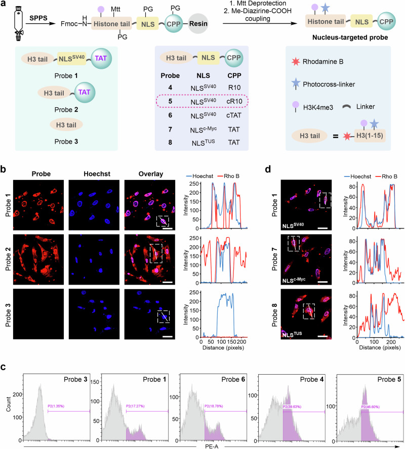
Fig. 2. Chemical synthesis and visualization of nucleus-targeted cell-permeable histone-tail-based photoaffinity probes.
a Synthetic scheme and structure of probes 1 to 8. b Confocal microscopy images of HeLa cells treated with probes 1–3 for 1 h at 37 °C. Probes 1–3 were visualized using Rho B fluorescence (red channel), and Hoechst 33258 was utilized for nuclear staining (blue channel). 2D intensity profiles corresponding to the lines displayed on the images (line scans) are depicted to the right of the micrographs. Scale bars: 20 μm. Confocal microscopy images shown in (b) are representative of independent biological replicates (n = 3). c Cellular uptake analysis of probes in HeLa cells by flow cytometry. Cells were treated with probes 1 and 3–6 for 30 min at 37 °C. d Confocal microscopy images and line scans of HeLa cells treated with probes 1, 7, and 8. Scale bars: 20 μm. Confocal microscopy images shown in (d) are representative of independent biological replicates (n = 2). Source data are provided as a Source Data file.

