
Figure 5.
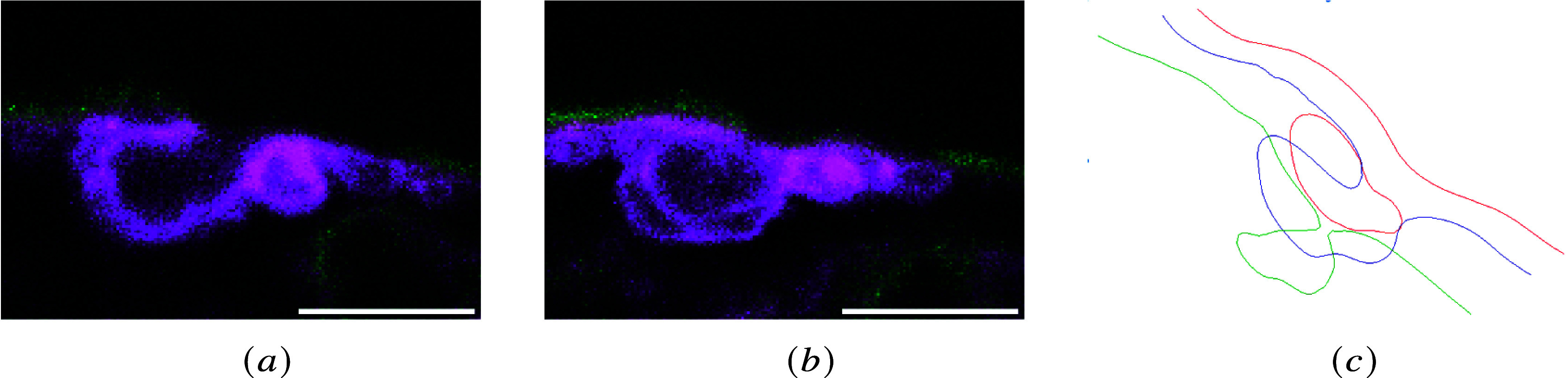
Different images from the same z-stack (the scale bar corresponds to
 ). (a) membrane profile corresponding to the luminal opening of the invagination of the luminal membrane and a circular cavity corresponding to a bleb; (b) membrane profile showing different cavities below the luminal membrane: the central cavity is a section of the ramified invagination, and the others beneath correspond to cytoplasmic voids; and (c) the cartoon shows a schematic representation of different longitudinal sections of the luminal membrane within the same z-stack.