
Figure 7.
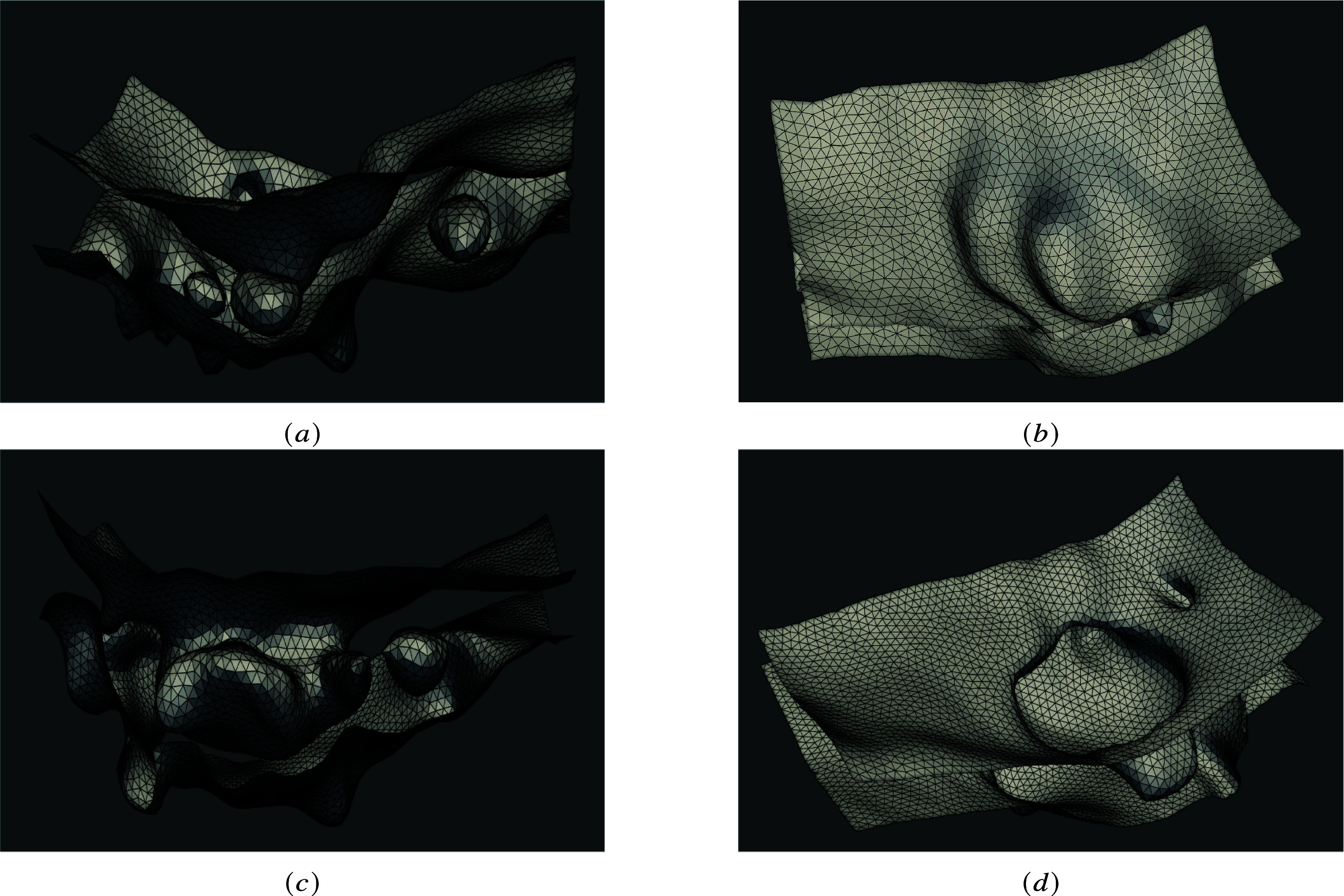
Membrane reconstruction. The meshes corresponding to different stages of the EHT are shown (90 min separate the acquisition timing between the top and the bottom panels). (a, c) a longitudinal view is shown, and we can observe the interior voids and blebs at the basal membrane and (b, d) the luminal part of the cellular membrane is shown, with its quite significant deepening and increased curvature at the rim (see (d)).