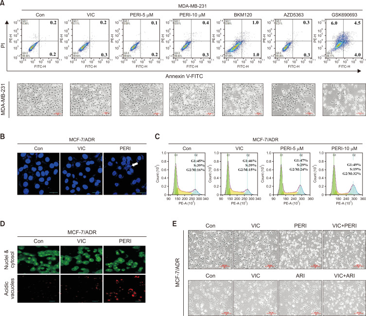
Fig. 3.
Perifosine increases DNA condensation, G2 arrest, and autophagy induction in P-gp overexpressing drug-resistant MCF-7/ADR cells. (A) MDA-MB-231 cells were treated with 5 nM VIC, 5 μM perifosine (PERI-5μM), 10 μM perifosine (PERI-10μM), 1 μM BKM120, 10 μM AZD5363, 10 μM GSK690693, or 0.1% DMSO (CON). After 1 day, all cells were also observed using an inverted microscope at ×40 magnification, and annexin V analyses were performed as described in Materials and Methods. (B) MCF-7/ADR cells were treated with 5 nM VIC, 10 μM perifosine (PERI), or 0.1% DMSO (CON). After 24 h, staining with DAPI was performed as described in Materials and Methods. (C) MCF-7/ADR cells were treated with 5 nM VIC, 5 μM perifosine (PERI-5μM), 10 μM perifosine (PERI-10μM), or 0.1% DMSO (CON). After 1 day, FACS analyses were performed as described in Materials and Methods. (D) MCF-7/ADR cells were treated with 5 nM VIC, 10 μM perifosine (PERI), or 0.1% DMSO (CON). After 24 h, staining with acridine orange was performed as described in Materials and Methods. (E) MCF-7/ADR cells were treated with 5 nM VIC, 5 μM perifosine (PERI), 5 μM aripiprazole (ARI), 5 nM VIC+5 μM perifosine (VIC+PERI), 5 nM VIC+5 μM aripiprazole (VIC+ARI), or 0.1% DMSO (CON). After 1 day, all cells were observed using an inverted microscope at ×40 magnification.

