Figure 5:

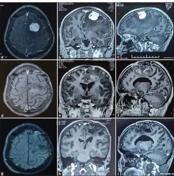
Illustrative case (a-c): Pre-operative contrast-enhanced magnetic resonance imaging (MRI) axial, sagittal, and coronal images of left frontal meningioma in a 20 year old male with seizure, headache, and right hemiparesis. The patient was operated on, and a near-total excision of the tumor was done. The histology report was suggestive of atypical meningioma. The patient received linear accelerator-based radiotherapy (60 Gy in 30 cycles) 3 months after surgery. (d-f): Follow Follow-up contrast MRI axial, sagittal, and coronal images obtained 8 months after surgery, showing few areas of ring enhancement noted in the right high frontal region – post-radiotherapy changes. (g-i): Follow-up contrast MRI axial, sagittal, and coronal images obtained 5 years after surgery showed a cystic lesion in the left frontal region with perilesional sclerosis associated with thinning of the overlying cortex suggestive of encephalomalacia and no evidence of residual or recurrent lesion noted.
