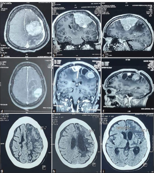
Figure 7:

Illustrative case (a-c): Pre-operative contrast enhanced magnetic resonance imaging (MRI) axial, sagittal, and coronal images of the left middle 1/3rd parasagittal meningioma in 35 years female with complex partial seizure, headache, right hemiparesis and slurring of speech. The patient underwent Simpson grade II excision of the tumor. The histology report was suggestive of secretory meningioma (WHO grades I). Seven years after surgery, the Patient came with complaints of right hemiparesis, seizure, and aphasia. (d-f): MRI axial, sagittal, and coronal images showed well defined extra-axial solid cystic lesion with septal enhancement in cystic lesion and homogeneous contrast enhancement in solid component along the left parasagittal region with surrounding mild intraparenchymal edema suggestive of recurrent meningioma. As the patient had poor pre-treatment factors for surgery, external beam radiotherapy was given (54 Gy in 30 cycles). (g-i): Post-radiotherapy non-contrast computed tomography head was suggestive of hypodensities in the left frontal and parietal regions with ex-vacuo dilatation of ipsilateral lateral ventricle and adjoining sulcal spaces suggestive of gliotic changes.
