. 2024 Dec 11. Online ahead of print. doi: 10.1039/d4lc00471j

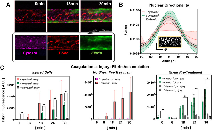
Fig. 2. Fluid shear stress and injury regulate coagulation levels in the PINCH devices. A) Confocal micrographs depicting accumulation of fluorescent fibrin (green) at PSer-positive (red) sites of vascular injury. Cytosolic dye (magenta) indicates uninjured cells. B) Nuclear directionality measurements reveal shear promotes more nuclear alignment along the direction of flow, 0° (n = 5). C) Pre-treatment of endothelium with fluid shear stress prior to injury limits subsequent coagulation (0 dyne cm−2 injury: N = 13; 0 dyne cm−2 no injury: N = 5; 5 dyne cm−2 injury: N = 6, 5 dyne cm−2 no injury: N = 5, 10 dyne cm−2 injury: N = 5, 10 dyne cm−2 no injury: N = 5; mean ± SEM, *p < 0.05, **p < 0.01, mixed-effects analysis). Greater coagulation levels are seen in injured vascular microchannels pre-treated with 0 and 5 dyne cm−2 shear stress as compared to 10 dyne cm−2.

Fig. 2