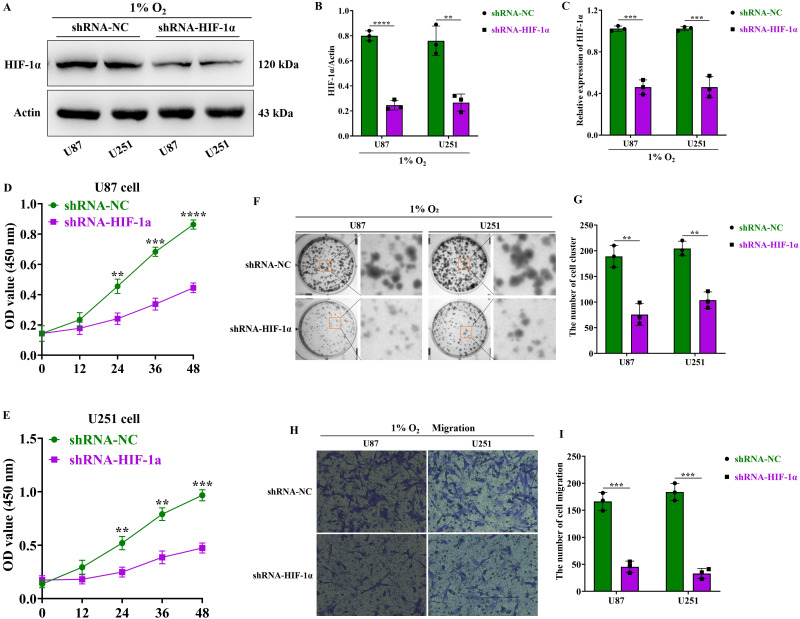
Figure 2.
HIF-1α promotes the malignant progression of GBM. (A) Western blot analysis of HIF-1α protein expression levels in GBM; (B) Graphical representation of HIF-1α protein expression levels; (C) Real-time quantitative PCR assessing HIF-1α mRNA expression in GBM. (D) CCK8 assay to measure U87 cell proliferation following HIF-1α knockdown; (E) CCK8 assay to measure U251 cell proliferation following HIF-1α knockdown. (F) Clonogenic assay to assess the colony-forming ability of U87 and U251 cells after HIF-1α knockdown; (G) Statistical analysis of the number of colonies formed. (H) Cell migration assay to evaluate U87 and U251 cell migration post-HIF-1α knockdown; (I) Statistical analysis of migrated cell numbers. Data are presented as mean ± standard deviation, and differences between two groups were analyzed using an unpaired t-test. **P <0.01, ***P <0.001, ****P <0.0001.

