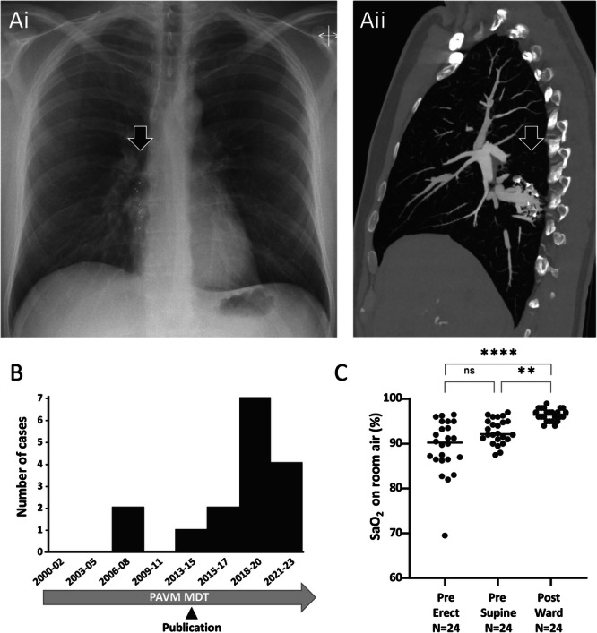
Figure 1.

(A) Complex right lower lobe PAVM following previous embolization with several Amplatzer vascular plugs. Preoperative images. (i) Chest x‐ray indicating Amplatzer plug radiopaque tips and additional soft tissue shadowing (arrowed). (ii) Sagittal images from a contrast enhanced thoracic CT scan demonstrating a persistent venous sac with multiple small residual feeding arteries, the diameter and position of which preclude complete embolization. (B) Case series: Serial number of elective thoracic surgical cases, noting date overseas audit of cumulative radiation doses in patients with PAVMs was published [4]. Cases from the third series (2023‐) are the subject of separate Case Reports with specific educational/training foci. (C) Pre and postoperative oxygen saturation (SaO2). SaO2 is a validated biomarker of right‐to‐left shunting [5], and did not differ between Series 1 and Series 2 pretreatment (means 89.2% vs. 89.6%). p values were calculated by Dunn's test post Friedman (**p < 0.01, ****p < 0.001).
