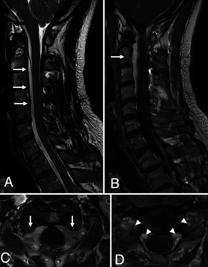
FIG. 2.

Preoperative cervical MRI. Sagittal T2-weighted MRI demonstrating an interdural cyst, starting from C2 and extending to the thoracic level (arrows, A). Note that no lesions were compressing the spinal cord. Sagittal postcontrast T1-weighted MRI demonstrating enhancement of the epidural venous plexus in the cervical region (arrow, B). Postcontrast axial T1-weighted MRI demonstrating enhancement of the epidural venous plexus at the C2 (arrows, C) and C4–5 (D) levels. Note the compression of the C5 nerve roots (arrowheads, D).
