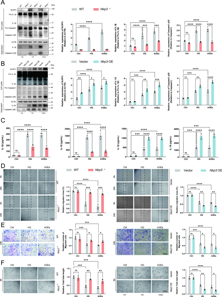
Fig. 4.
Regulated angiogenesis function of EPC by NLRP3 inflammasome in response to HG or AGEs. (A) Western blot analysis of NLRP3, Caspase1, and IL-1β cleavage in EPC from WT or Nlrp3−/− mice following stimulation with HG (30 mM) or AGEs (200 µg/ml) for 72 h. The supernatant was collected directly after a 72-hour incubation period with high glucose and AGEs. (B) EPC from wild-type mice were transfected with Vector or Nlrp3. Western blot analysis of NLRP3, Caspase1, and IL-1β cleavage in EPC transfected with Vector or Nlrp3 following stimulation with HG (30 mM) or AGEs (200 µg/ml) for 72 h. (C) ELISA analysis of IL-18, and IL-1β in supernatants of EPC from WT or Nlrp3−/− mice and EPC transfected with Vector or Nlrp3 following stimulation with HG (30 mM) or AGEs (200 µg/ml) for 72 h. (D) Wound healing of EPC response to HG (30 mM) or AGEs (200 µg/ml) in vitro. (E) Migration assay with EPC treated with HG (30 mM) or AGEs (200 µg/ml) was performed. (F) The angiogenic function of EPC was evaluated by tube formation assay. *p < 0.05, **p < 0.01, ***p < 0.001, ****p < 0.0001. Representative results from three biologically independent experiments

