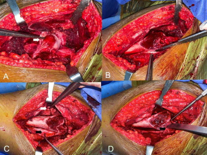
Fig. 2.
A) L-shaped anterior retinacular incision (white arrow) in stable SCFE. B) Subperiosteal dissection with a scalpel was made for the posterolateral retinacular flap. C) Anterior (white arrow) and posterior retinacular (black arrow) flaps. D) To complete the separation of the posterolateral flap, the small remaining bony chip of the posterolateral stable trochanter (Black arrow) was separated using a straight osteotome through the cut surface of trochanter

