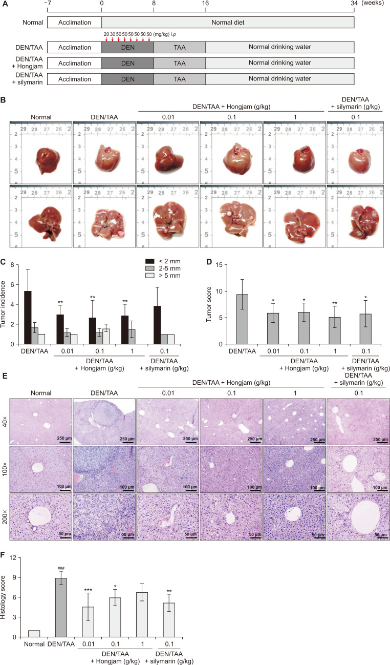
Figure 1. Effect of Hongjam supplementation on tumor incidence and tissue lesion in the DEN/TAA-induced HCC model.
(A) Schematic representation of the experimental protocol for the DEN/TAA-induced HCC model. DEN was administered intraperitoneally for 8 weeks, followed by TAA in drinking water for an additional 8 weeks. Animals were sacrificed at week 34. (B) Representative images of extracted livers. (C) Tumor count and size measurements. (D) Tumor scoring based on size: tumors < 2 mm were scored as 1 point, those 2-5 mm as 2 points, and tumors > 5 mm as 5 points. (E) H&E staining showing the effect of Hongjam on tissue lesions (magnification: ×40, ×100, ×200). (F) Quantification of H&E staining intensity using histopathological scoring. Statistical significance was determined using Tukey’s multiple comparison test, with data presented as mean ± SD (n = 8). ###P < 0.001 vs. Normal group; ***P < 0.001, **P < 0.01, *P < 0.05 vs. DEN/TAA group. DEN, diethylnitrosamine; TAA, thioacetamide; HCC, hepatocellular carcinoma; H&E, hematoxylin and eosin; i.p., intraperitoneally.

