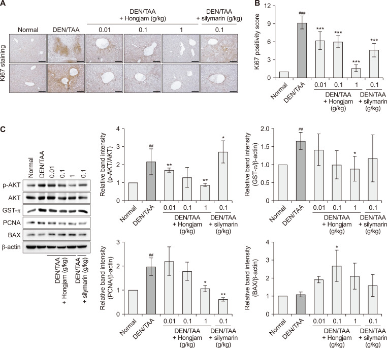
Figure 3. Hongjam suppresses proliferation in the DEN/TAA-induced HCC model.
(A) IHC staining of Ki67 in liver tissue, indicating levels of cell proliferation (magnification: × 100). (B) Quantitative analysis of Ki67 positivity in IHC staining (magnification: ×100; scale bar: 100 μm). (C) Western blot analysis of liver-derived proteins showing the expression levels of p-AKT, AKT, GST-π, PCNA, and BAX. Statistical significance was determined using a Tukey’s multiple comparison test. Data are presented as mean ± SD (n = 3). ###P < 0.001, ##P < 0.01 vs. Normal group; ***P < 0.00, **P < 0.01, *P < 0.05 vs. DEN/TAA group. DEN, diethylnitrosamine; TAA, thioacetamide; HCC, hepatocellular carcinoma; IHC, immunohistochemistry; p-AKT, phosphorylation of protein kinase B; GST-π, glutathione S-transferase π; PCNA, proliferating cell nuclear antigen; BAX, Bcl-2-associated X protein.

