. 2025 Jan 8;13:RP98661. doi: 10.7554/eLife.98661

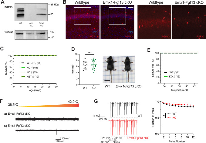
Figure 2. Excitatory neuronal knockout of Fgf13 does not result in premature death and seizure susceptibility.

(A) Western blot shows partial loss of Fgf13 from Emx1-Fgf13 cKO hippocampal tissue, compared to full knockout in Nestin-Fgf13 cKO hippocampus. Vinculin used as a loading control. (B) Fluorescent immunohistochemistry of hippocampal tissue validates Fgf13 knockout (scale bar, 100 μm). (C) Emx1-Fgf13 cKO mutant mice survive past 1 month of age (log-rank test, p=ns). (D). Body mass at postnatal day 14 (P14) shows that Emx1-Fgf13 cKO are not different in size (scale bar, 2 cm) (t-test, p=ns). (E) Emx1-Fgf13 cKO are not susceptible to hyperthermia induced seizures (log-rank test, p=ns). (F) EEG recordings during hyperthermia protocol show Emx1-Fgf13 cKO do not exhibit heat-induced seizures. (G) Emx1-Fgf13 cKO neurons exhibit diminished long-term inactivation (two-way ANOVA, *, p<0.05), though the deficit is not sufficient to cause seizures (WT, N=2, n=15; KO, N=2, n=15). Example traces for WT and Emx1-Fgf13 cKO neurons are shown on the left.

Figure 2—source data 1. Original TIFF files saved from Bio-Rad Gel doc system for gels shown in Figure 2A, together with a PDF file identifying the respective portions displayed.

Figure 2.

Figure 2—figure supplement 1. Excitatory neuronal knockout of Fgf13 results in loss of FGF13-S.
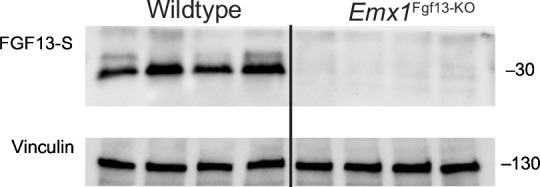
Figure 2—figure supplement 1.

Western blot performed with FGF13-S-specific antibody. Vinculin used as a loading control.
Figure 2—figure supplement 1—source data 1. Original TIFF files saved from Bio-Rad Gel doc system for gels shown in Figure 2—figure supplement 1, together with a PDF file identifying the respective portions displayed.
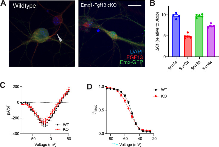
Figure 2—figure supplement 2. Excitatory neuronal knockout of Fgf13 does not result in sodium current deficits.

Figure 2—figure supplement 2.

(A) Primary hippocampal neuron cultures were generated from Emx1-Cre wildtype (left) or Emx1-Fgf13 cKO male mice, and Emx1 +excitatory cells were labeled by infection with a Cre-dependent AAV8-DIO-GFP virus. Immunocytochemistry was performed with a pan-FGF13 antibody (scale bar, 20 μm). Arrow indicates FGF13 in the axon initial segment, absent in the Emx1-Fgf13 cKO neurons. (B) Reverse transcriptase quantitative polymerase chain reaction of neuronal voltage-gated sodium channels shows levels of expression for specific channels relative to Actb (N=4–5 mice). (C) Emx1-Fgf13 cKO neurons do not exhibit differences in macroscopic sodium currents (two-way ANOVA, p=ns). Peak current-voltage (I–V) curves shown; WT N=3 mice, n=18 cells; KO N=4 mice, n=18 cells. (D) Emx1-Fgf13 cKO neurons do not have a significant difference in steady-state inactivation (V1/2 WT=−52.04 [95% CI, −53.60 to -50.47]; V1/2 KO = –54.47 [95% CI, –56.4––52.54]). WT N=3 n=21; KO N=4 n=37.