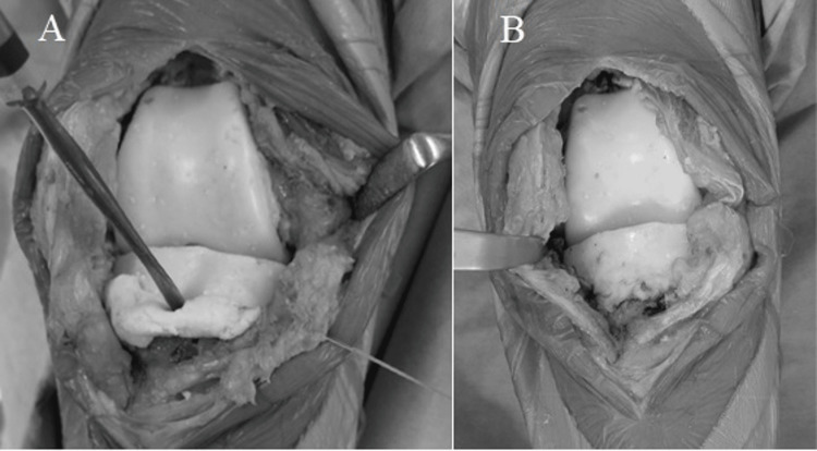
Figure 4. A photograph of the tibial side cement spacer insertion technique.
Additional cement containing antibiotics was used below the tibial mold to fill the gap space at the knee extension position (A). The appearance after the cement spacer was placed, leaving minimal joint gap, is shown in (B).

