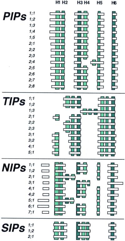
Figure 3.
Schematic structure of MIP encoding genes in Arabidopsis. Horizontal bars and gaps depict exons and intron positions, respectively. Parts encoding transmembrane helices H1 to H6 according to an alignment with GlpF are indicated by vertical bars. The color on the vertical bars shows homologous transmembrane helices in the first and second halves of the MIPs. The exons and transmembrane helices are drawn to scale but the positions of helices are schematic. Helices encoded on two exons are only indicated on the exon where the major part is encoded. Small indels in the alignment of different genes, positioned between two helices on the same exon, are not shown.

