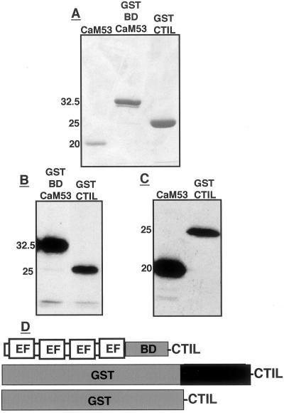
Figure 5.
CaM53, GST-BDCaM53, and GST-CTIL protein substrates and their prenylation. A, Stained SDS-PAGE of the purified protein substrates. B, Fluorogram showing prenylation of GST-BDCaM53 and GST-CTIL fusion recombinant protein. C, Fluorogram showing prenylation of CaM53 and GST-CTIL recombinant proteins. Numbers in A through C denote molecular mass in kilodaltons. D, Schematic presentation of CaM53, GST-BDCaM53, and GST-CTIL. BD, Basic domain; EF, Ca2+-binding EF hands.

