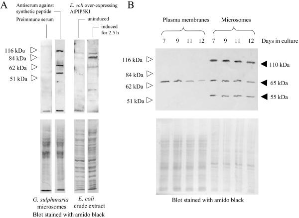
Figure 2.
A putative 65-kD PtdInsP kinase protein decreases in the plasma membrane during the stationary phase. Antisera were produced in rabbits against a synthetic 15-amino acid peptide representing a conserved PtdInsP 5 kinase domain from Arabidopsis. A, Equal protein amounts (20 μg) of microsomes from G. sulphuraria were separated by SDS-PAGE and blotted to PVDF membranes. Immunodetection with rabbit pre-immune serum, and with anti-PtdInsP kinase antiserum, as indicated. Soluble protein extracts from E. coli expressing AtPIP5KI were separated by SDS-PAGE and blotted to PVDF membranes. Immunodetection with anti-PtdInsP kinase antiserum with extract from uninduced cells, or from cells induced for 2.5 h with 1 mm IPTG, as indicated. Blots stained with amido black are shown (lower). B, Plasma membranes and microsomes were prepared from G. sulphuraria samples harvested over the period of time following the transition from logarithmic growth to the stationary phase. Equal amounts of protein (20 μg) were separated by SDS-PAGE and blotted to PVDF membranes. Upper, Immunodetection of PtdInsP kinase protein with the anti-PtdInsP kinase antiserum. Lower, Blot stained with amido black. White arrows indicate Mr markers.

