Abstract
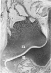
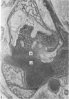
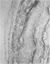
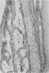
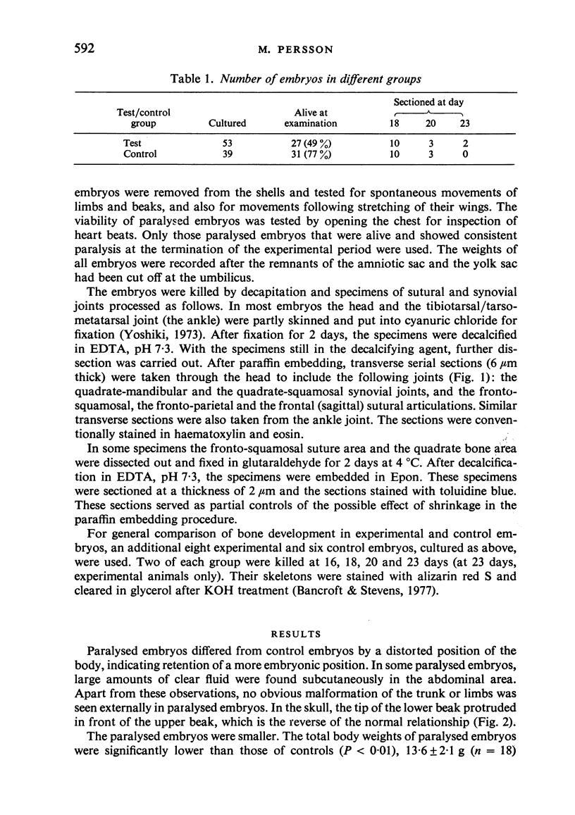
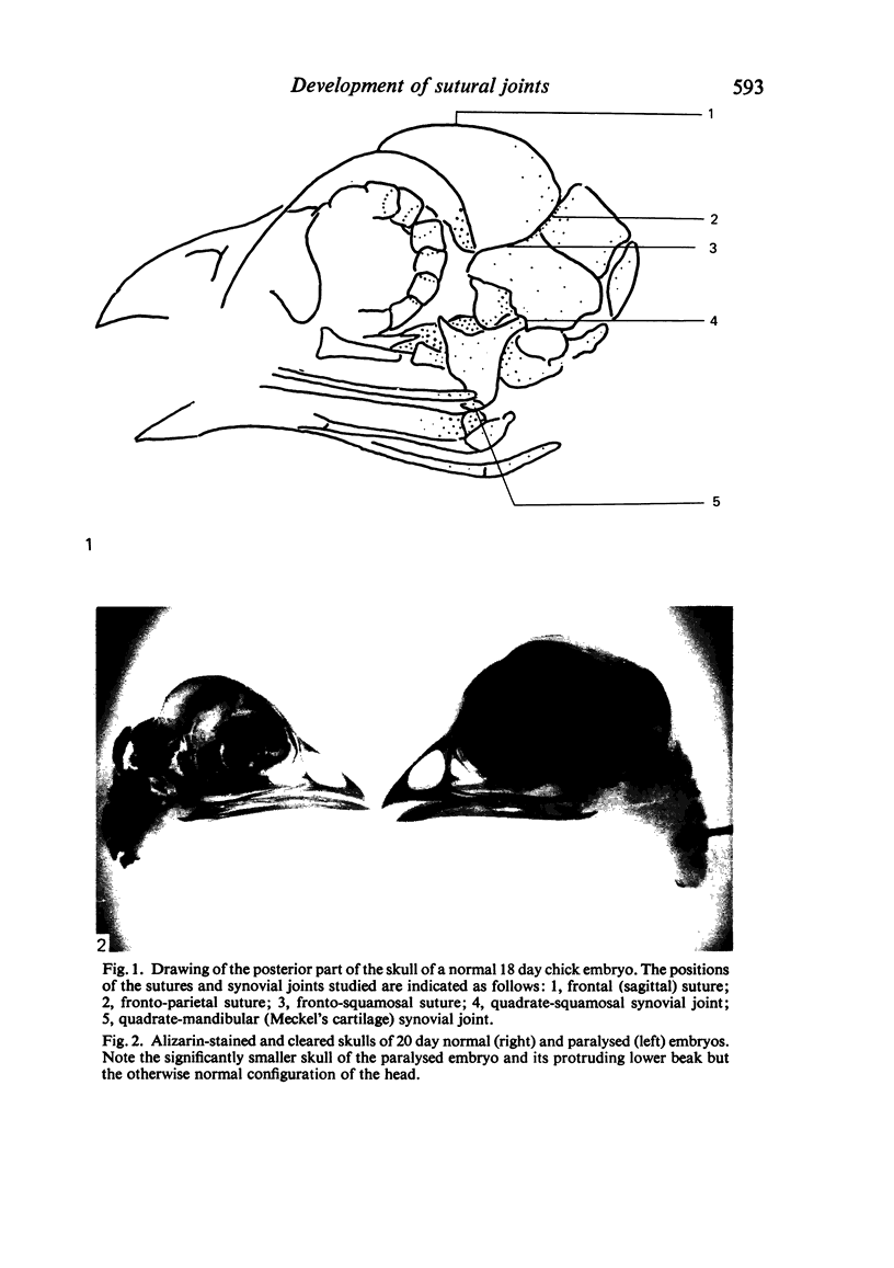
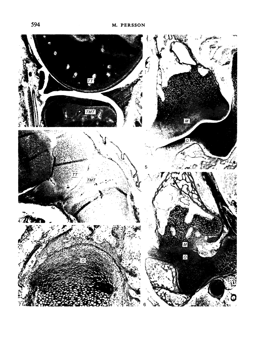
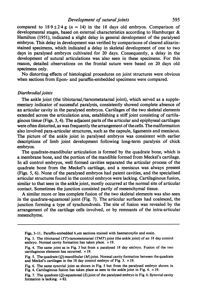
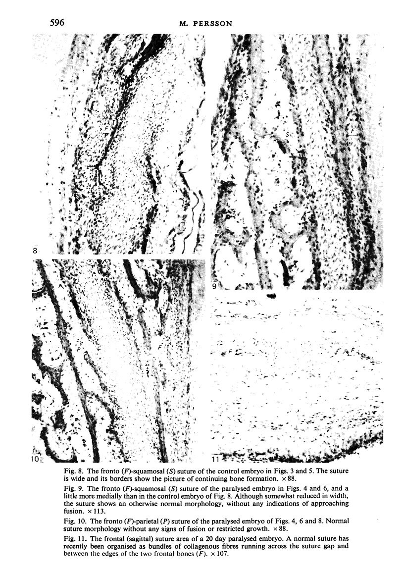
Chick embryos were paralysed in ovo with a neuromuscular blocking agent between 8 and 20 days of incubation. To evaluate the rôle of muscular activity in the development of sutural articulations, sutures of the cranial vault of control and paralysed embryos were studied histologically and the findings compared with the effect of the agent on the development of the ankle joint and some synovial joints of the jaws. Paralysed embryos showed a consistent lack of development of joint cavities in synovial joints. In most embryos, fusion of opposing cartilaginous elements had occurred. In contrast to synovial joints, sutural articulation showed a micromorphology comparable to that of controls. The findings indicate that different embryonic factors regulate the development of sutural and synovial articulations. Movements of neuromuscular origin play no essential role in the morphogenetic development of sutures, but are a prerequisite for the formation of joint cavities and other specialised structures of synovial joints.
Full text
PDF
Images in this article
Selected References
These references are in PubMed. This may not be the complete list of references from this article.
- DRACHMAN D. B., COULOMBRE A. J. Experimental clubfoot and arthrogryposis multiplex congenita. Lancet. 1962 Sep 15;2(7255):523–526. doi: 10.1016/s0140-6736(62)90399-9. [DOI] [PubMed] [Google Scholar]
- Hall B. K. A simple, single-injection method for inducing long-term paralysis in embryonic chicks, and preliminary observations on growth of the tibia. Anat Rec. 1975 Apr;181(4):767–777. doi: 10.1002/ar.1091810408. [DOI] [PubMed] [Google Scholar]
- LELKES G. Experiments in vitro on the role of movement in the development of joints. J Embryol Exp Morphol. 1958 Jun;6(2):183–186. [PubMed] [Google Scholar]
- Moss M. L. Functional anatomy of cranial synostosis. Childs Brain. 1975;1(1):22–33. doi: 10.1159/000119554. [DOI] [PubMed] [Google Scholar]
- Murray P. D., Drachman D. B. The role of movement in the development of joints and related structures: the head and neck in the chick embryo. J Embryol Exp Morphol. 1969 Nov;22(3):349–371. [PubMed] [Google Scholar]
- Oppenheim R. W., Pittman R., Gray M., Maderdrut J. L. Embryonic behavior, hatching and neuromuscular development in the chick following a transient reduction of spontaneous motility and sensory input by neuromuscular blocking agents. J Comp Neurol. 1978 Jun 1;179(3):619–640. doi: 10.1002/cne.901790310. [DOI] [PubMed] [Google Scholar]
- PRITCHARD J. J., SCOTT J. H., GIRGIS F. G. The structure and development of cranial and facial sutures. J Anat. 1956 Jan;90(1):73–86. [PMC free article] [PubMed] [Google Scholar]
- Persson K. M., Roy W. A., Persing J. A., Rodeheaver G. T., Winn H. R. Craniofacial growth following experimental craniosynostosis and craniectomy in rabbits. J Neurosurg. 1979 Feb;50(2):187–197. doi: 10.3171/jns.1979.50.2.0187. [DOI] [PubMed] [Google Scholar]
- Persson M., Roy W. Suture development and bony fusion in the fetal rabbit palate. Arch Oral Biol. 1979;24(4):283–291. doi: 10.1016/0003-9969(79)90090-6. [DOI] [PubMed] [Google Scholar]
- Smith D. W., Töndury G. Origin of the calvaria and its sutures. Am J Dis Child. 1978 Jul;132(7):662–666. doi: 10.1001/archpedi.1978.02120320022004. [DOI] [PubMed] [Google Scholar]
- Ten Cate A. R., Freeman E., Dickinson J. B. Sutural development: structure and its response to rapid expansion. Am J Orthod. 1977 Jun;71(6):622–636. doi: 10.1016/0002-9416(77)90279-2. [DOI] [PubMed] [Google Scholar]
- Yoshiki S. A simple histological method for identification of osteoid matrix in decalcified bone. Stain Technol. 1973 Sep;48(5):233–238. doi: 10.3109/10520297309116630. [DOI] [PubMed] [Google Scholar]


