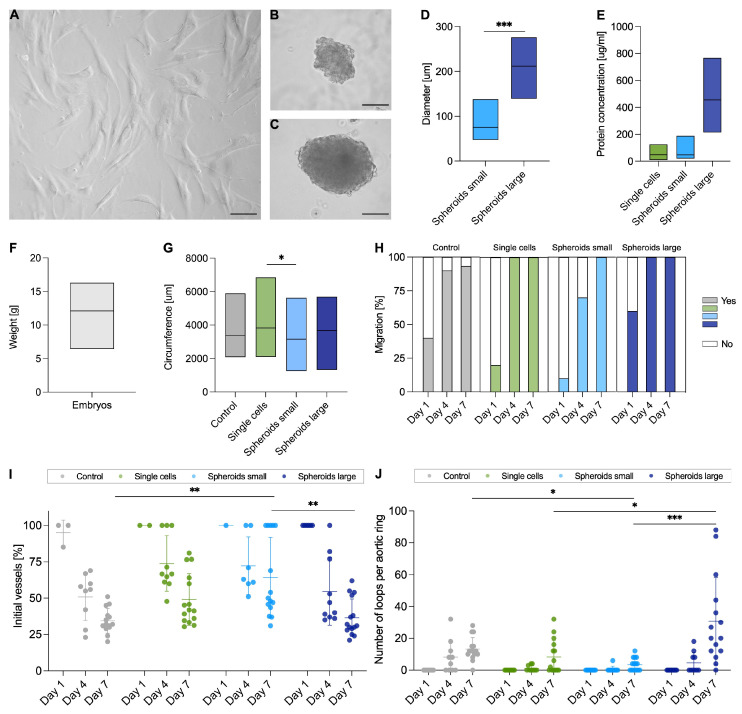
Figure 2.
Collecting of secretomes, comparability of chicken aortas, and pattern analysis. For the production of the secretomes, human adipose-derived mesenchymal stem cells were cultivated either as single cells ((A); scale bar 100 μm) or as spheroids small ((B); scale bar 50 μm) and spheroids large ((C); scale bar 50 μm). After 3 days, the diameter of the spheroids was determined (D). The protein concentration of the harvested secretomes was measured (E). Before harvesting the aorta, each chicken embryo was weighed (F) and for the aortic rings used in the aortic ring assay, the circumference was measured under the microscope (G). Migration of the aortic rings was determined on days 1, 4, and 7, and visible vessel sprouting was considered positive (Yes; (H)). Vessel architecture was assessed by primary initial vessels in relation to total vessel number (I) and by the number of loops formed (J). The aortic rings were treated with either serum-free medium (Control), secretomes of single cells, spheroids small or spheroids large (G–J). Data are shown as boxplots with min to max values and mean (D–G). Groups were compared using Mann–Whitney test (D), one-way ANOVA with Tukey’s multiple comparison tests (G), and nonparametric Kruskal–Wallis test for day 7 time point (I,J); * p < 0.05; ** p < 0.01; *** p < 0.001.

