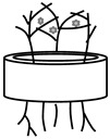
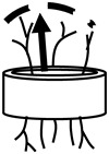
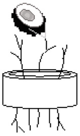
Table A1.
Characterization of neovascular phenotype in ex vivo culture based on categories. The column synonyms refer to terms that were used synonymously with the respective parameters in the cited studies.
| Category | Parameter | Synonyms | Definition | Evaluation | Benefits | Disadvantages | References |
|---|---|---|---|---|---|---|---|
Explant
|
Outgrowth | n/a | Migration of progenitor cells and response to culture condition | Number of explants with a neovascular outgrowth over total explants | Quick overview of the quality of explants and matrices, an early indicator for technical problems throughout the experiment |
The significance of the parameter decreases with a small number of samples | [102,103] |
| Area | n/a | Area (A) of the projection of the aortic ring into the image | A in mm2 equals the number of pixels that form the object multiplied by a calibration constant square | A has almost the same value for each set of experiments and may serve for standardization | Software (e.g., ImageJ) for the evaluation and for the generation of binary images is necessary, A depends on the age and size of the animal |
[44] | |
| Circumference | Size | Length of the line that delimits the vessel | Measurement of the outer perimeter of the vessel from captured images | The vessels develop exclusively at the cutting edge of the explant, explant circumference can be used for normalization |
Depends on the age and size of the animal | [104,105] | |
| Shape | Form | Shape factor (F) of arterial ring | Perimeter2/4πA describes the deviation of an object from a true circle (F), it gives a minimal value of 1 for a circle and larger values for shapes having a higher ratio of perimeter to area |
F has almost the same value for each set of experiments, results do not depend on the geometry of aortic rings |
Software (e.g., ImageJ) for the evaluation and for the generation of binary images is necessary | [44,105] | |
Pattern
|
Mesh | Cellular organization, morphology | Quantification of cellular organization, which are closed areas delineated by segments and associated junctions | Use of extension of “Angiogenesis Analyzer” plugin for ImageJ software, results for mean size of meshes and total mesh area |
Suitable as an early marker, as over time, segment interruptions resulting in mesh fusions increase | Software-based evaluation of the vectorial objects through corresponding algorithms, formed network often varies in different physiological and pathophysiological environments |
[16,95,106] |
| Structure | Tree detection, branching pattern, microvessel distribution |
Geometry of microvessels as a function of distance to the aortic ring, or as a skeletal illustration |
Tree modeling consists of segmentation followed by skeletonization of vascular structures, number of intersections of microvessels with a grid defined as the successive boundaries of the dilated aortic ring |
Both qualitative (skeletonization) and quantitative (microvessel distribution) evaluation is possible | Manual evaluation is not feasible, qualitative evaluation is based on a subjective comparison of the images by eye and shows junctions without counting the branches, avoid shadows due to phase contrast lighting and small acellular structures during segmentation |
[16,44,46] | |
| Angle | Degree of branching vessel | Angle between branches | Measurement of value in degrees of how two branches relate to each other | Provides information about vascular patterns, network formation, and parallel alignment of vessels | Software-based (e.g., Synedra View) evaluation is needed | [95] | |
| Loop | Endothelial network loop, loop formation, vessel lumen |
Loops with at least three sides | Count of endothelial network loops | Quantification identifies effects on the development of endothelial networks | A cell counter tool is recommended, inadequate or uneven polymerization of the matrix, as well as its damage, can interfere with successful network development |
[8,106,107] | |
| Network properties 
|
Vessel area | Sprouting area, network area, vascular area outgrowth area, tubule area |
Total area quantifies the surface of all measured vessels | Based on the measured length and width of vessels, the area can be calculated, the value can be given as a total value per area or region or as a percentage, as well as a relative value, whereby the vessel area at time T = 0 is subtracted from the vessel area after further corresponding treatment days |
Vessel area increases as the vascular network grows, the relative vessel area enables comparison over time |
Use of image processing software (e.g., IKOSA, Wimasis) as the measurement of the surface area of all vessels cannot be determined by manual analysis, new vascular sprouts formed during angiogenesis are thin and may not affect the total vessel area as much as other parameters |
[1,8,46,106,108,109,110,111,112] |
| Radial outgrowth | Radial network growth, circumference of the arterial ring, aortic ring area, sprouting area, maximal distance migrated, area of migrated vessels, microvessel outgrowth |
Distance from the aortic ring to the furthest distal outgrowth of cells | Measuring the distance from the cut end of the aortic segment to the approximate mean point of vessel growth | Value correlates with the apparent number of cells forming vessels, same images can be used to quantify network properties, including radial outgrowth and loop formation in one assay quadrant |
Dependent on the growing pattern, less informative value than the total vessel length |
[8,44,103,110,113,114,115,116,117,118] | |
| Vessel length | Sprout length, tubule length, microvessel length, length of capillary-like microtube, segment length, branch length |
Length of vessel centerline | Determination of the length of individual vessels, including their branches, in an image using a software application, values can be expressed as the sum of all lengths (total vessel length), the average vessel length, the average length of segments, which includes the distance between junctions, and the maximum vessel length |
Vessel elongation is a critical process during angiogenesis, the total length of the vascular network is influenced by the appearance of new branches and the elongation of existing vessels, widely used parameter with a high informative value, vessel length is an important parameter for measuring the effects of exogenous factors on angiogenesis, length of vascular tree is associated with a later stage of development |
Difficult to analyze with high vessel density and overlapping of vessels, many studies focus on assessing the larger vessels while paying little attention to microvessels, vessel length can be affected by factors such as cell adhesion or chemoattractant gradients |
[1,5,8,11,16,44,46,95,103,104,105,106,108,110,111,112,115,118,119,120,121,122,123,124] | |
| Vessel thickness | Vessel diameter, width vessel, wall thickening |
Diameter of a vessel | Determine thickness by measuring the diameter of a vessel | Changes in the mean thickness of the vasculature are related to angiogenic processes, vessel width reveals primary and secondary orders, leading to the identification of main and minor vessels of the vascular network |
Decrease in the mean diameter of the vascular network through the development of new thin sprouts | [1,46,95,108,109] | |
| Speed | n/a | The ratio of distance covered to time spent | Measurement over time of prominent vessel elongation in living culture | Examination of vascular proliferation possible | Not suitable for a low degree of vascular development | [107] | |
Sprouting
|
Vessel counts | Vessel density, number of microvessels, tube number, number of capillaries |
Cells are organized as vessels after endothelial-driven cells sprouting from the aortic ring, and the number of vessels is counted accordingly, total vessel number based on branch and terminal points counting |
Documentation by phase contrast microscopy followed by manual or computer-assisted counting of vessels (per field of view or per ring), values can be displayed as the total number of vessels (vessel counts), vessel density (total number of vessels per area), or the number of vessels per aortic ring |
Widely used parameter with a high informative value, number of vessels is an important parameter for measuring the effects of exogenous factors on angiogenesis, the potency of angiogenic activity can be assessed by the number and growth rate of newly formed vessels and calculated as an angiogenic score (vessel density in relation to distance from the ring) |
Difficult and time-consuming when counted manually, comparability between studies may be difficult due to different ways of presenting the values, accurate quantification based on visual counts may not be possible, as the number of neovessels and the complexity of the vascular networks in the culture may be very high |
[1,5,11,44,95,102,103,105,106,107,108,110,118,119,120,122,123,124,125] |
| Branch | Sprout, segment |
Line that is either connected to the main vessel by a junction or is located between two junctions | Counting the number of branching in vessels (manually or computer-assisted) | New branches develop by sprouting from pre-existing vessels, the number of branches provides information on the way the vessels are organized and develop in the presence of stimulators or inhibitors of angiogenesis |
No minimum inclusion criteria exist on how many aligned cells a branch must consist of (e.g., three or more cells), no distinction is made in the manual evaluation as to whether extremities or connections within the vascular tree are involved |
[1,11,44,46,95,105,106,108,113,118,120,123] | |
| Junction | Branching point, node, intersection |
Junction consists of a minimal structure and allows branching in a skeletonized network, junction is determined where it has at least three adjacent branches |
Counting the number of junctions, the values can be presented as the total number of junctions per ring or per field of view, as the average number of junctions per vessel, or as the ratio average number of junctions per average number of branches |
It allows a statement on the degree of branching and thus on the architecture of the vessel tree, manual as well as computer-assisted evaluation is possible |
In case of overlapping of vessels, an evaluation can no longer be performed accurately, and significance loses its validity, manual count of junctions is very time-intensive |
[8,16,106,123] | |
| Break-away capillaries |
n/a | The foremost segments of the sprouts can detach from the parent microvessels and migrate as isolated break-away capillaries on the advancing front of the outgrowth | Quantitation of break-away capillaries based on light microscopic images | This effect is particularly evident in a mid- or long-term culture, a microvessel culture with a small number of break-away capillaries shows thicker and more mature capillaries |
No intensively studied parameter in the in vitro setting so far, data available for collagen gel culture of rat aorta |
[119] | |
Cellular level
|
Endothelial cells | ECs | Flattened cell type that forms a layer covering blood vessels | Staining of cells without or with aortic ring (e.g., GFP, Dil-Ac-LDL) prior to the experiment for in vitro cell tracking possible or afterward for identification of this specific cell type (e.g., CD31, vWF, BSL-B4, BSL-1), analysis by fluorescence microscopy (including capturing Z-series image stacks), calculating the number of ECs aligned in vessels, an area with the maximal sprouting, or number of branches |
Additional evaluation of further parameters that characterize the structure of the vascular network, e.g., β-catenin interacts in ECs, and its loss may reduce junction stability followed by increased vascular permeability, closer examination of tip and trunk cells possible, the aortic ring sprouts anatomically mimic microvessels in vivo, making them suitable for studies of paracrine signaling between endothelial cells, pericytes, and smooth muscle cells |
Fibroblasts may first have to be removed by a short digestion of the sample, after transfection, the expression of the exogenous gene often decreases at the time when its presence could be relevant for the morphological changes in the cells, after transduction and depending on the titres used, cells may show an altered morphological pattern of sprouting and vascular formation, in contrast to retroviruses, adenoviruses stress the cell through the transcription and translation of adenoviral genes, the range of antibodies available depends on the species to be investigated |
[3,44,110,113,116,124,126,127,128] |
| Fibroblast-like cells | Pericytes, smooth muscle cells, myofibroblasts |
Capillary-like structures with isolated fibroblast-like cells, primarily confined in peri- aortic location, spatially isolated contractile cells on capillaries, play an important role in the main- tenance of vascular and tissue homeostasis |
Specific staining enables the determination of the cell type through the exclusion criterion, selectively uptake of fluorescent Dil-Ac-LDL by endothelial cells, remaining fibroblast-like cells are detected as unstained, counting the number of pericytes covering a 100-um-long stretch of neovessel |
Myofibroblast-endothelial interactions can be modeled (transduction of EC with a mCherry retroviral vector, and myofibroblasts with a GFP retroviral vector), GFP-labeled pericytes can be utilized to track motility and the recruitment of pericytes into the EC tubes can be observed explant assays are considered to come closest to mimicking the in vivo situation because they include the surrounding nonendothelial cells (such as smooth muscle cells and pericytes) and a supporting matrix, the EC tubes in EC pericyte co-cultures become much narrower and more elongated compared to EC-only tubes, which become wider and less long over time |
Pericyte antibody panel is not available for every species | [3,8,37,44,129,130,131,132] |
Key: A: Area; BSL-1: Griffonia (Bandeiraea) Simplicifolia Lectin I; BSL-4: Griffonia Simplicifolia Lectin I (GSL I) Isolectin B4; Dil-Ac-LDL: 1,10-Dioctadecyl-3,3,30,30-tetramethyl-indocarbocyanine perchlorate; EC: Endothelial cell; F: Factor shape; GFP: Green fluorescent protein; n/a: Not appropriate; vWF: Von Willebrand Factor.
