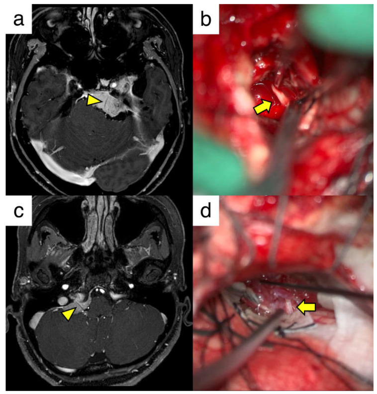
Figure 2.
Illustrative cases of skull base meningiomas with cranial nerve penetration. (a) T1-weighted image with contrast of a patient with a left petroclival meningioma. The yellow arrowhead shows the contrast defect in the abducens nerve. (b) Intraoperative view of the abducens nerve penetrating the tumor (yellow arrow). (c) T1-weighted image showing the contrast of the patient with a right petrous meningioma. The yellow arrowhead indicates a contrast defect in the lower cranial nerve. (d) Intraoperative view of the glossopharyngeal nerve penetrating through the tumor (yellow arrow).

