Figure 2.
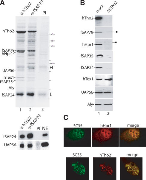
Identification of the human THO complex and its colocalization with splicing factors in nuclear speckles. (A) Immunoprecipitations from RNase-treated HeLa nuclear extracts using anti-hTho2 antibody (lane 1), anti-fSAP79 antibody (lane 2), and preimmune serum (lane 3). (H) Heavy chain; (L) light chain. Proteins were identified by mass spectrometry. Bands indicated by the fish icon are, from top to bottom, RAD 50 homolog, SAP 145, CDC27 homolog, and NMT 55. A dash indicates molecular weight markers, from top to bottom (in kilodaltons): 181, 115, 82, 63, 48, 37, and 26. (Lower panel) Western analysis of immunoprecipitated samples. (NE) Nuclear extract. (B) Western analysis of mock- and hTho2-immunodepleted nuclear extracts. The square indicates nonspecific bands. (C) Immunofluorescence of HeLa cells. Merge shows coimmunofluorescence with the SC35 antibody.

