Figure 5.
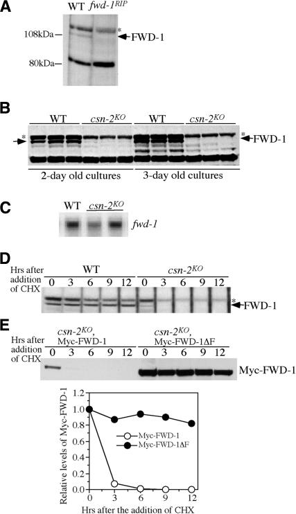
The FWD-1 levels are drastically reduced in the csn-2 mutant, probably due to its rapid autoubiquitination-mediated degradation. (A) Western blot analysis showing the expression of FWD-1 in the wild-type and fwd-1RIP strains. The arrow indicates the FWD-1 specific band in the wild-type strain. The asterisk indicates an unspecific cross-reacted protein band recognized by our FWD-1 antiserum. (B) The expression of FWD-1 in the wild-type and csn-2KO strains after 2 or 3 d of growth in liquid medium. (C) Northern blot analysis showing the expression of fwd-1 mRNA in the wild-type and csn-2KO strains. (D) Western blot analysis showing the degradation of FWD-1 after the addition of CHX. Similar results were obtained in three independent experiments. (E) Degradation of Myc-FWD-1 or Myc-FWD-1ΔF (lacking its F-box) expressed in the csn-2 mutants after the addition of CHX. The densitometric analysis of the results is shown in the bottom panel.

