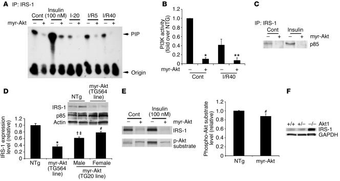
Figure 2.
IRS-1/PI3K signaling is inhibited in myr-Akt hearts. (A) IRS-1–associated PI3K activity was analyzed in the hearts perfused without (Cont, n = 5) or with insulin (100 nM, 10 minutes; n = 3) or subjected to 20 minutes of ischemia (I-20) followed by 5 minutes or 40 minutes of reperfusion. PIP, phosphoinositide; origin, initial position of the reaction mixer. (B) Quantitation of mean spot density from cumulative data are shown for control (n = 5 each) and ischemia/reperfusion (n = 3 each) normalized to the values for the control-perfused NTg hearts. *P < 0.001 versus control-perfused NTg; **P < 0.01 versus NTg after ischemia/reperfusion. (C) Lysates from the hearts with or without insulin stimulation were immunoprecipitated with IRS-1 and immunoblotted for the p85 subunit of PI3K. Representative data from 4 independent experiments are shown. (D) Representative immunoblot for IRS-1 and p85 from cardiac lysates of myr-Akt (TG564 line) (n = 6) or NTg (n = 5) mice. Actin was included as a loading control. Densitometric quantitation from the different line of myr-Akt–Tg (TG20 line, male and female; n = 3 each) in addition to TG564 line is shown in the bar graph. *P < 0.001 versus NTg; †P < 0.01 versus NTg and TG564; ‡P < 0.02 versus TG20 female; #P < 0.05 versus NTg. (E) Lysates from the hearts with or without insulin stimulation were immunoblotted first with anti–IRS-1 antibody and subsequently with anti–phospho-Akt substrate antibody. Representative data from 4 independent experiments are shown. Densitometric quantitation from control hearts is shown in the bar graph. (F) Representative immunoblots of IRS-1 from cardiac lysates of Akt1+/+, Akt1+/–, and Akt1–/– mice are shown from 3 independent experiments. GAPDH immunoblotting is shown as a loading control.

