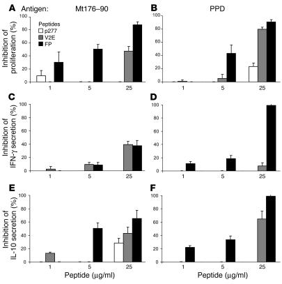
Figure 3.
FP inhibits the T cell response to Mt. Lymph node cells from Mt-immunized rats were activated in vitro with Mt176–190 (A, C, and E) or PPD (B, D, and F) in the presence of FP, V2E, or p277. Proliferation (A and B), IFN-γ secretion (C and D), and IL-10 secretion (E and F) were assayed. The data are presented as mean inhibitions ± SD (n = 3 or more). The uninhibited T cell proliferative responses were 12258 ± 578 cpm and 1488 ± 103 cpm for PPD and Mt176–190, respectively. The background proliferation in the absence of antigen (8–10 days after a previous activation by incubation with APC and PPD or the Mt176–190 peptide) was 380 ± 15 cpm. IFN-γ secretion was 1155 ± 254 pg/ml and 1289 ± 310 pg/ml for cells activated by PPD and Mt176–190, respectively. IL-10 secretion was 364 ± 69 pg/ml and 314 ± 23 pg/ml for cells activated by PPD and Mt176–190, respectively.

