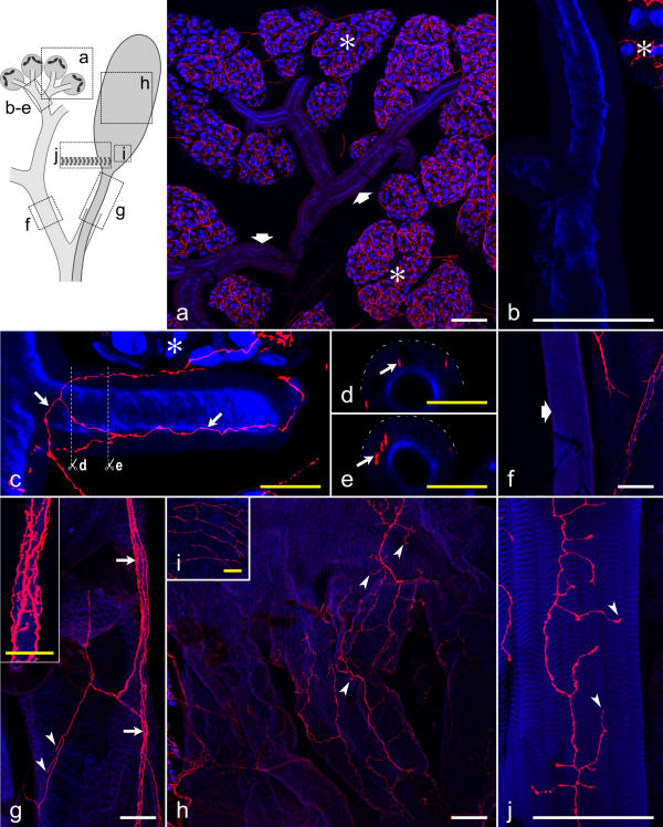
Figure 10.
Distribution of serotonergic fibers on salivary ducts, the reservoir, and the reservoir muscle The upper left inset indicates the structures shown in a-j. a-c, f-j: Summarized views of confocal image stacks through whole-mounts, double-labeled with anti-serotonin (red) and BODIPY FL phallacidin (blue). Asterisks in a,b,c indicate acinar tissue. a: A dense network of serotonergic fibers is associated with the acini (asterisks), whereas small salivary ducts (broad arrows) are mostly without serotonergic fibers. b: A small salivary duct without serotonergic innervation at higher magnification. c: A small salivary duct with a network of serotonergic fibers (arrows). d,e: Vertical sections through the salivary duct shown in c (planes indicated by white lines), demonstrating that the serotonergic fibers (arrows) reside below the duct surface (broken lines). f: A large salivary duct (broad arrow) without serotonergic innervation. g: The reservoir duct is accompanied by a nerve (arrows and inset) containing several serotonergic fibers. Fibers in a superficial position within the nerve have numerous varicosities (inset). Individual fibers also extend over the reservoir duct and have terminals (arrowheads) associated with this structure. h: A loose network of serotonergic fibers, with their terminals (arrowheads), covers the middle part of the reservoir. i: At the orifice, the reservoir has a relatively dense network of serotonergic fibers on its surface. Note that i is enlarged twofold compared with h. j: The reservoir muscle contains numerous serotonergic fiber terminals (arrowheads). White scale bars = 100 μm; yellow scale bars = 25 μm

