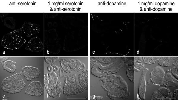
Figure 2.
Specificity of anti-serotonin and anti-dopamine labeling
a-d: Fluorescence confocal images, representing the summarized view of 9-μm-thick image stacks. e-h: Nomarski contrast images of the same areas. a,b: Cryostat sections of salivary glands incubated with anti-serotonin in the absence or in the presence of 1 mg/ml serotonin. c,d: Sections reacted with anti-dopamine in the absence or in the presence of 1 mg/ml dopamine. Immunoreactivity of the tissue is highly reduced in the presence of the corresponding antigen. Scale bars = 100 μm

