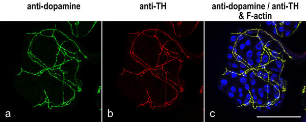
Figure 3.
Codistribution of anti-dopamine and anti-TH immunolabeling Whole-mounts of salivary glands were triple-labeled with anti-dopamine (green), anti-TH (red) and BODIPY FL phallacidin (blue), and imaged by confocal microscopy. The image shows a lobule of acinar tissue; the peripheral cells are arranged in pairs and their apical arrays of phallotoxin-stained microvilli appear as "bow ties". A sparse network of fibers resides on the tissue and is labeled by both anti-dopamine and anti-TH. Scale bar = 100 μm

