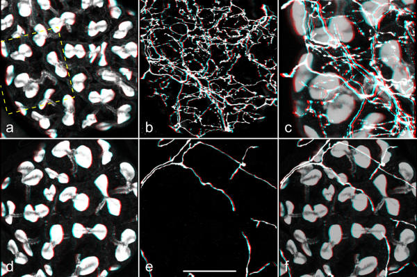
Figure 8.
Three-dimensional (red-green) views of serotonergic and dopaminergic fibers associated with acinar lobules Salivary glands were double-labeled with BODIPY FL phallacidin (a,d) and anti-serotonin (b) or anti-dopamine (e). Stacks of confocal images were recorded, and three-dimensional reconstructions were made by using Carl Zeiss LSM510 software. c,f: The corresponding images of staining with phallotoxin and with antibody were added (a+b or d+e; the phallotoxin image was multiplied with the factor 0.7 to reduce its intensity) in order to present both staining patterns together. The rectangle in a indicates the area that is presented at higher magnification in c. b,c: A dense network of serotonergic fibers extends throughout the entire acinar tissue. e,f: Dopaminergic fibers, in contrast, form a loose network only on the acinar surface. Scale bar = 50 μm

