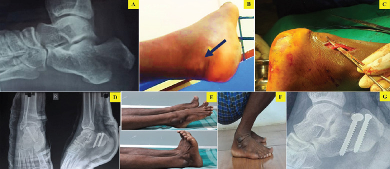
Figure 2.

(A) pre-operative X-ray, (B) pre-operative clinical picture showing the skin impalement (black arrow), (C) intraoperative picture showing sural nerve identification and preservation, (D) post-operative X-ray showing cannulated cancellous screw fixation, (E-G) range of movements and X-ray after union at final follow-up.
