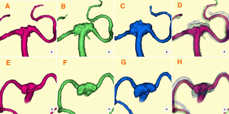
Figure 2. Serial 3D silent MRA images capturing the progression of RC enlargement.
A, E) Fourteen days post-treatment: The RC formed and extended superoposteriorly, as shown in anterior-posterior (A) and superior-inferior (E) projections. B, F) Six months post-treatment: The RC expanded postero-laterally (horizontally), as depicted in anterior-posterior (B) and superior-inferior (F) projections. C, G) One year post-treatment: The RC exhibited continued postero-lateral (horizontal) growth, along with an additional minor superior (vertical) extension at its central region, as observed in anterior-posterior (C) and superior-inferior (G) projections. D, H) Fused images overlaying time-series 3D silent MRA data provide a comprehensive visualization of the progressive RC enlargement, shown in anterior-posterior (D) and superior-inferior (H) projections.
MRA: magnetic resonance angiography; RC: residual cavity

