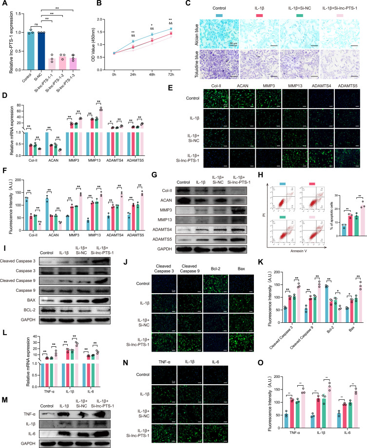
Figure 3.
The deficiency of lnc-PTS-1 exacerbated ECM catabolism, apoptosis, and inflammatory responses in chondrocytes. (A) qRT-PCR analysis of lnc-PTS-1 expression of C28/I2 cells transfected with siRNAs or negative control (n=3). (B) CCK-8 assays were performed to determine the cell viability of transfected C28/I2 cells under IL-1β treatment at 24, 48, and 72 h (*P < 0.05, **P < 0.01, Control vs IL-1β; &&P < 0.01, IL-1β+Si-NC vs IL-1β+Si-lnc-PTS-1) (n=6). (C) Alcian blue and Toluidine blue staining (scale bar: 50 μm) were conducted to determine the ECM anabolism of transfected C28/I2 cells under IL-1β treatment (n=3). (D) qRT-PCR, (E and F) representative IF images and quantitative analysis, and (G) Western Blot analysis showed expression levels of ECM metabolism indicators of lnc-PTS-1 knock down C28/I2 cells with IL-1β (n=3). (H) The apoptosis rate of C28/I2 cells was showed by flow cytometry (n=3). (I) Western Blot analysis, and (J and K) representative IF images (scale bar: 25 μm) and quantitative analysis showed apoptosis-related indicators expression levels of lnc-PTS-1 knock down C28/I2 cells with IL-1β (n=3). (L) qRT-PCR, (M) Western Blot analysis, and (N and O) representative IF images (scale bar: 25 μm) and quantitative analysis showed expression levels of inflammatory factors of lnc-PTS-1 knock down C28/I2 cells with IL-1β (n=3). Data are presented as mean ± SD, Statistical analysis was performed using one-way analysis of variance (ANOVA); *P < 0.05, **P < 0.01, ns, non-significance, P ≥ 0.05.

