
Fig. 2.
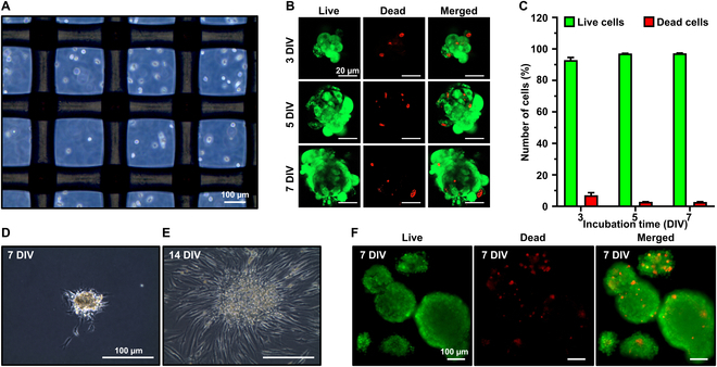
Fabrication of HFDPC spheroids and cell viability evaluation. (A) Optical microscopy immediately after seeding. (B) Live/dead assay and (C) the ratio of live and dead cells at 3, 5, and 7 DIV. HFDPC spheroids cultured on TCP at (D) 7 DIV and (E) 14 DIV. (F) Live/dead assay on the HFDPC spheroids prepared by hanging drop.