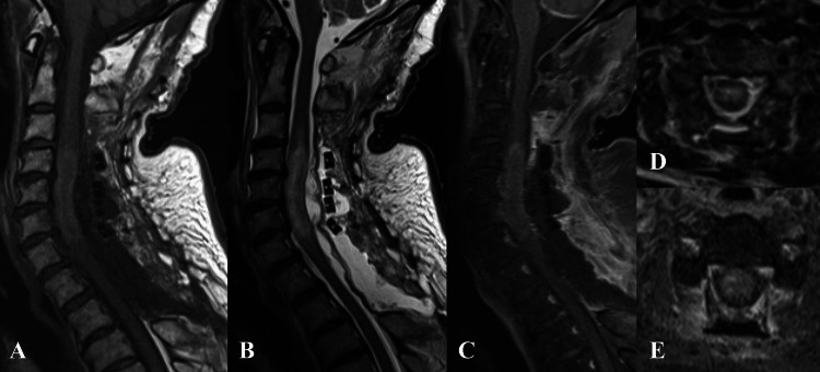
Figure 4. Postoperative gadolinium-enhanced MRI of the cervical spine.
The dural sac is decently expanded, but the intradural signal change at the C4-Th1 level resides (arrowhead).
Aː Sagittal T1 fat-saturated weighted image; Bː Sagittal T2-weighted image; Cː Sagittal gadolinium-enhanced image; D: Axial T2-weighted image at the C4/5 level; E: Axial gadolinium-enhanced image at the C4/5 level

