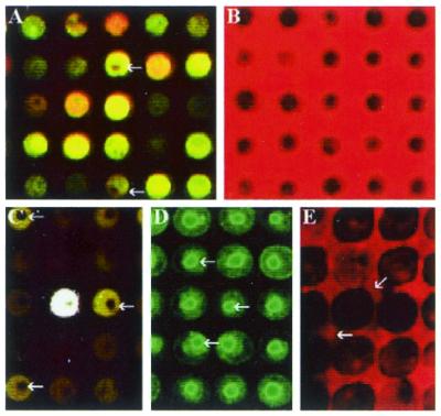
Figure 4.
Images of various DNA spots. (A) A small set of typically labeled DNA spots. The arrow indicates a small unlabeled portion in the center of a spot. (B) DNA spots before the hybridization procedure. The DNA appears as a black color. (C) The arrows point to typical ‘donut’ spots. This is an area within the spot where there is no hybridization. (D) The penetration of the probe towards the center of the spots as shown by the arrows. (E) The diffusion of DNA from the spots into the hybridization solution.

