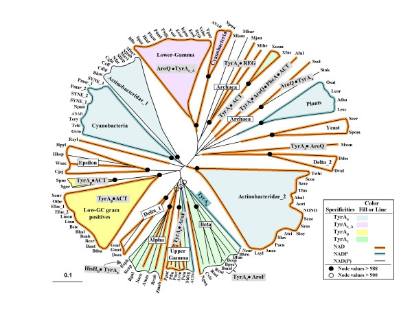
Figure 2.

Phylogenetic tree for trimmed core domains of selected members of the TyrA Superfamily. Acronyms used for the various organisms are given in alphabetical order in Table 2. (A more extensive listing that includes organisms not shown in Fig. 2 and which also is hyperlinked to all of the individual GenBank records is given in Table S1. A similar table that also includes compilation of known and predicted substrate specificities is maintained at AroPath [73]. Lineages possessing experimentally established TyrAa, TyrAp, TyrAc or TyrAc_Δ proteins are indicated by fill colors specified in the legend. Three specificity patterns for the pyridine nucleotide substrate are shown by line colors (see figure box). Although the cyanobacteria are depicted as having NADP+-specific TyrA proteins, some of them can also accept NAD, albeit to a lesser degree. All proteins having an aspartate residue homologous to D-32 of the E. coli NADTyrAc_Δ domain are presumed specific for NAD+. Fusion of TyrA domains with other catalytic domains is indicated within grey boxes (AroQ•TyrA, TyrA•AroF, HisHb•TyrA, and TyrA•AroQ•PheA•ACT) using the convention of a bullet to represent the interdomain area. The boxes overlap any relevant lineages. TyrA proteins having carboxy-terminal fusions with regulatory domains (TyrA•ACT and TyrA•REG) are also shown. The distance scale bar at the bottom left represents substitutions per site.
