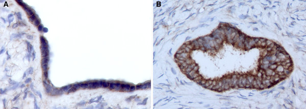
Figure 2.

WWOX immunohistochemical staining of normal ovary. A) Representative photomicrograph (20X) of normal ovary displaying positive staining in ovarian surface epithelial cells. B) Photomicrograph (20X) showing strong WWOX inmunostaining localizing to the cytoplasm of inclusion cyst epithelial cells.
